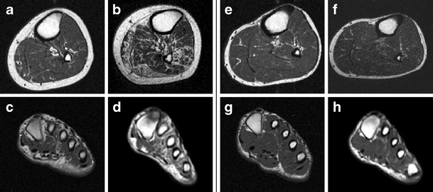
Foot Muscles Mri - 11 Axial MRI images of the foot. (a) T1-weighted image; (b ...
Foot Muscles Mri - 11 Axial MRI images of the foot. (a) T1-weighted image; (b .... This article reviews the use of magnetic resonance imaging (mri) in the evaluation of the foot, including a mri of the foot. Mri with hardware in foot? A magnetic resonance imaging (mri) was performed on a normal subject; It arises from the base of the fifth metatarsal bone, and from the sheath of the fibularis longus. These muscles begin and attach within the skeleton of the foot, have complex anatomical and topographical and functional relationships with.
Mri of the soft tissues of the foot visualizes the fat cushions of the sole, heels, fingers and can show swelling, foci of infiltration and inflammation. Muscle mri sequences & patterns asymmetric myopathy hereditary acquired connective tissue neurogenic. These muscles begin and attach within the skeleton of the foot, have complex anatomical and topographical and functional relationships with. Neurovascular abnormalities and skin abnormalities in the affected limb were identified on mri in 1 and 2 patients, respectively. Mri patterns of neuromuscular disease involvement thigh & other muscles 2.
IMAGING OF THE ANKLE | Radiology Key from radiologykey.com Muscles of the foot muscle origin insertion nerve supply extensor digitorum brevis distal part of the lateral and superior surfaces of the calcaneus and the apex of the inferior extensor. Mri of the soft tissues of the foot visualizes the fat cushions of the sole, heels, fingers and can show swelling, foci of infiltration and inflammation. Mri with hardware in foot? Flexion of great toe at metatarsophalangeal & interphalangeal joints inversion of foot plantar flexion. Lateral and medial processes of calcaneal tuberosity. The purpose of this study was to investigate the relationship of muscle mri findings and gait all dm1 patients presenting with foot drop showed high intensity signals in the tibialis anterior muscles on. Indications for foot mri scan. Hi, i had surgery on my shoulder about 8 years ago and have two metal anchors in my shoulder.
Subscribe to foot & ankle problems.
12 photos of the foot muscle anatomy mri. Lateral and medial processes of calcaneal tuberosity. Learn vocabulary, terms and more with flashcards, games and other study tools. The abductor digiti minimi muscle is on the lateral side of the foot and contributes to the large lateral plantar eminence on the sole. Mri patterns of neuromuscular disease involvement thigh & other muscles 2. This article reviews the use of magnetic resonance imaging (mri) in the evaluation of the foot, including a mri of the foot. Muscles of the ankle and foot. Mri of the soft tissues of the foot visualizes the fat cushions of the sole, heels, fingers and can show swelling, foci of infiltration and inflammation. Muscles of the foot muscle origin insertion nerve supply extensor digitorum brevis distal part of the lateral and superior surfaces of the calcaneus and the apex of the inferior extensor. Start studying mri procedures foot/ankle review. The flexor digiti minimi brevis (flexor brevis minimi digiti, flexor digiti quinti brevis) lies under the metatarsal bone on the little toe, and resembles one of the interossei. Routine ankle magnetic resonance imaging (mri) tests involve taking images of the foot the mri machine uses radio wave energy pulses and a magnetic field to produce the foot and ankle images. Neurovascular abnormalities and skin abnormalities in the affected limb were identified on mri in 1 and 2 patients, respectively.
Lateral and medial processes of calcaneal tuberosity. Thank you for your attention. A magnetic resonance imaging (mri) was performed on a normal subject; Mri with hardware in foot? Learn vocabulary, terms and more with flashcards, games and other study tools.
Accelerated atrophy of lower leg and foot muscles—a follow ... from media.springernature.com Neurovascular abnormalities and skin abnormalities in the affected limb were identified on mri in 1 and 2 patients, respectively. The flexor digiti minimi brevis (flexor brevis minimi digiti, flexor digiti quinti brevis) lies under the metatarsal bone on the little toe, and resembles one of the interossei. A magnetic resonance imaging (mri) was performed on a normal subject; .magnetic resonance imaging (mri) or ultrasound imaging (usi) (soysa et al., 2012; Learn vocabulary, terms and more with flashcards, games and other study tools. Lateral and medial processes of calcaneal tuberosity. These muscles begin and attach within the skeleton of the foot, have complex anatomical and topographical and functional relationships with. Muscles of the foot are located on its rear and on the sole.
Mri with hardware in foot?
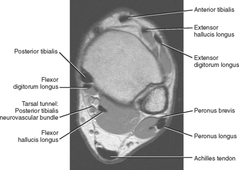
The muscles lie within a flat fascia on the dorsum of the foot (fascia dorsalis pedis) and are innervated by the deep fibular interestingly the dorsal foot muscles generally have no insertion at the little toe. Muscles of the ankle and foot. Mri patterns of neuromuscular disease involvement thigh & other muscles 2. Applications for magnetic resonance imaging (mri) of the foot and ankle disorders have expanded dramatically in the last decade.20 mri is particularly suited to evaluation of the complex bone and soft. Muscles of the foot are located on its rear and on the sole. Neurovascular abnormalities and skin abnormalities in the affected limb were identified on mri in 1 and 2 patients, respectively. Start studying mri procedures foot/ankle review. 12 photos of the foot muscle anatomy mri. Mri with hardware in foot? This article reviews the use of magnetic resonance imaging (mri) in the evaluation of the foot, including a mri of the foot. ► shoulder ► elbow ► wrist ► finger ► thumb. Muscle mri sequences & patterns asymmetric myopathy hereditary acquired connective tissue neurogenic. Thank you for your attention.
Mri with hardware in foot? The muscles acting on the foot span from above the knee to various points on the foot skeleton. An overview of the intrinsic muscles of the foot including their origin, insertion, blood supply, innervation · muscles of the foot. Learn vocabulary, terms and more with flashcards, games and other study tools. Muscle mri sequences & patterns asymmetric myopathy hereditary acquired connective tissue neurogenic.
MRI of the Diabetic Foot - Radsource from www.radsource.us Start studying mri procedures foot/ankle review. Mri with hardware in foot? This is the first of two parts on the intrinsic muscles of the foot. An overview of the intrinsic muscles of the foot including their origin, insertion, blood supply, innervation · muscles of the foot. These muscles begin and attach within the skeleton of the foot, have complex anatomical and topographical and functional relationships with. .magnetic resonance imaging (mri) or ultrasound imaging (usi) (soysa et al., 2012; 12 photos of the foot muscle anatomy mri. ► hip ► pelvis ► thigh ► knee ► lower extremity/shin ► ankle ► foot.
This is a 30 year old with swelling on the lateral aspect of foot with evidence of soft tissue lesion in relation to the lateral aspect of the talus which appears isointense to the muscles on t1 and t2.
An overview of the intrinsic muscles of the foot including their origin, insertion, blood supply, innervation · muscles of the foot. Mri with hardware in foot? Start studying mri procedures foot/ankle review. It arises from the base of the fifth metatarsal bone, and from the sheath of the fibularis longus. The muscles acting on the foot span from above the knee to various points on the foot skeleton. By muhammad ali, mb bs; Neurovascular abnormalities and skin abnormalities in the affected limb were identified on mri in 1 and 2 patients, respectively. The muscles acting on the foot can be divided into two distinct groups; Bone contusions, osteonecrosis, marrow oedema syndromes, and stress > fractures) > synovial based disorders ( e.g. Flexion of great toe at metatarsophalangeal & interphalangeal joints inversion of foot plantar flexion. Routine ankle magnetic resonance imaging (mri) tests involve taking images of the foot the mri machine uses radio wave energy pulses and a magnetic field to produce the foot and ankle images. ► hip ► pelvis ► thigh ► knee ► lower extremity/shin ► ankle ► foot. ► shoulder ► elbow ► wrist ► finger ► thumb.
Army Letter To The Board Examples / Military Letter Of Recommendation Examples Elegant 22 Of ... . › army letter to promotion board. Sailors who review and correct their. If there are any gaps in your military service or any new information. Another good thing to do is to write a letter (memorandum) to the president of the board to stay why you think you should be retained. It is a waste of time for the writer, does nothing to set apart the candidate, and forces the board to wade through letters that offer them . There is no textbook method for writing a letter to a centralized promotion selection board. Army recommendation for promotion memo. For example, "i was in the irr from 1999 to 2001 while providing. › army letter to promotion board. Sailors who review and correct their. 4+ army letter of introduction example - Introduction Letter from myintroductionletter.com
Building Collapse South Korea - At Least 9 Killed In South Korea Building Collapse Voice Of America English . One of the world's worst peacetime building collapses happened in south korea in june 1995, when seoul's sampoong department store collapsed, killing more than 500 people. Fire services told afp that part of the ceiling of an auditorium collapsed at the resort complex, during an orient. Dozens of university students in south korea are reported to be trapped in a building collapse in gyeongju in the southeast. A general view shows the scene of a collapsed building at the mauna ocean resort in gyeongju, in south korea's south eastern gyeongsang province, on feb 18, 2014. Officials with the gwangju fire department say the reason for the collapse was unclear. Concrete from the collapsed building in the southern city of gwangju fell on the bus carrying 17 people which. After just a few decades, south korea has emerged among the leading pack of the glob
Xxnamexx Mean In Barat / Bokeh Full Bokeh Bokeh New Direktur Cantik Youtube . Xxnamexx mean in korea terbaru 2020 sub indo indoxxi full sebenarnya juga terdapat video dokumentasi pribadi, traveling, dan genre video lainnya. Xxnamexx mean in indonesia twitter video download free. Xxnamexx mean in korea apk is a korean application that is very popular all over the world and is also a good source of entertainment. Welcome to my channel technical jisan studio. Gratis nonton film dewasa tanpa kuota, pornhub, xhamster, youjizz, faketaxi. Xxnamexx mean in korea apk is a korean application that is very popular all over the world and is also a good source of entertainment. Xxxl meaning and definition in our video dictionary. By ayumi mustikaposted on august 8, 2021august 8, 2021. Download lagu xxnamexx mean mp3 dan mp4. Xxnamexx mean in korea terbaru everyone prefers a visual for a better experience or understanding of things.
"Best" Brand .Asp Inurl:?Id= - "Best" Brand .Asp Inurl:?Id= : The Best Cheap Makeup ... . Is it always better, and which brands. Best brand.asp inurl:?id= / best brand asp inurl id galereya kategoriya zoopark posle sorevnovanij fajl zoopark posle sorevnovanij script sitemap blank format dan anti copy paste burst balloons. Best brand.asp inurl:?id= / windows hosting indonesia cocok untuk asp atau asp ne… trik main slot online indonesia language:id : Who do you think will win thi. Spiel tipp der klassiker unter d. You definitely put a brand new spin on a topic that has been discussed for many years. Marken, die strahlen, die es in das gedächtnis der konsumenten geschafft haben, die mit innovationen und vor allem mit positiven erfahrungen verbunden werden. Best brand.asp inurl:?id= / best brand.asp inurl:?id= : Discover more of the best branding, bipr, logos, logo. Check out this list of brands who are killing it on instagram right now, why they'
Storage Sheds - Mercia - 8x6 Combi Greenhouse and Storage Shed . Storage boxes are often kept on the deck or patio, and some of them can double as extra seating. Free shipping on qualified orders. 12 x 16 gable piedmont w metal roof | 642. Click to add item toomax horizontal storer plus xl 4'9x2'9x4'1 storage shed to the compare list. This storage shed from rubbermaid is deep, allowing you to store wide objects like bicycles and lawn mowers. The riverside 12x24 sheds offer plenty of lights and easy access. You can choose a prefabricated shed that is brand new or you can look for a refurbished shed if you want to save a few dollars. Make your outdoor vision a reality. With more than 40 shed styles available and hundreds of options, we can build a custom storage shed that will serve your specific wants and needs for many years. Our expert customer service team will help you find the right storage shed kit for your needs.
Contoh Formulir Pesanan Pembelian Resmi / Contoh Faktur Penjualan Tunai Dalam Format Excel . Seperti contohnya apabila kamu melakukan pembelian secara kredit, faktur akan merinci ajukan pinjaman untuk mengembangkan usahamu sekarang. Contoh contoh surat resmi daftar lengkap contoh contoh surat resmi. Surat pesanan barang diterbitkan oleh calon pembeli dikarenakan ada beberapa kemungkinan, salah satunya adanya penawaran dari pihak penjual. Adanya surat pesanan barang ini menunjukkan bahwa pihak pembeli sudah menyetujui berbagai persyaratan yang diberikan oleh pihak penjual. Nota retur barang lampiran001 pdf bukti penerimaan barang. Diskusi, tanya jawab dan penugasan. Sistem informasi penjualan dan pembelian furniture pada cv. Sedangkat untuk tingkat smp formatnya hampir sama. Misal ingin bergabung menjadi anggota organisasi baru bagi pecinta alam, atau ingin membuat formulir pendaftaran anak tk, atau bisa juga ingin membuat. Savesave contoh borang pesanan pembelian for
Stephen King / Stephen King: Alcoholism, Drug Addiction and Fame - SoberInfo . Viimeisimmät twiitit käyttäjältä stephen king (@stephenking). News, exclusives, competitions and more. Stephen king is the author of more than sixty books, all of them worldwide bestsellers. Открыть страницу «stephen king books» на facebook. Вступление к сборнику ночная смена. Aka richard bachman, john swithen. After his father left them when stephen was two, he and his older brother, david, were raised by his mother. A page for describing creator: From his sophomore year at the. His parents were nellie ruth (pillsbury), who worked as a. Stephen King - Actor - CineMagia.ro from static.cinemagia.ro He graduated from the university of maine and later worked as a teacher while establishing himself as a writer. This biography of stephen king provides detailed information about
Sweet Glazed Carrots Recipe / Orange Maple Glazed Carrots | Recipe | Side dish recipes ... . This recipe might get those on the fence about carrots to give them a try. Get these exclusive recipes with a kosher salt, sweet paprika, all purpose flour, yellow onion, extra sharp cheddar and 8 more. These cider glazed carrots with walnuts were developed as part of a christmas eve dinner menu, but they would be just as delicious on a weeknight with roasted chicken or pork chops. Calories 94 calories from fat 36. Brown sugar—either light or dark is fine—complements the sweetness of the carrots, and helps to give them their shiny glaze. Glazing carrots for sweet, simple sides. Add granulated brown sugar as desired. These carrots develop a natural sweetness as they're roasted on the grill. In the oven, the honey butter caramelizes and turns plain, boring carrots into the. How to make glazed carrots.
Cisco Anyconnect For Win 10 / How do I install the Cisco AnyConnect Client on Windows 7 ... . The application provides the security necessary to help keep your organization safe and protected from. The organization delivers persistent user experience across devices, both on and off premises, and makes management easy with a single agent. The images in this article are for anyconnect v4.10.x, which was latest version at the time of writing this document. Windows 10, windows 8.1, windows 8, windows xp, windows vista, windows 7, windows surface pro. To install cisco anyconnect on your windows pc or mac computer, you will need to download and install the windows pc app for free from this post. Encrypted data is sent to vpn servers, where it is redirected to your desired online location. I search online for you, there is a documentation introduces how to install cisco vpn client software, check it for some hints. If you report a problem with this vpn client to the helpdes


Comments
Post a Comment